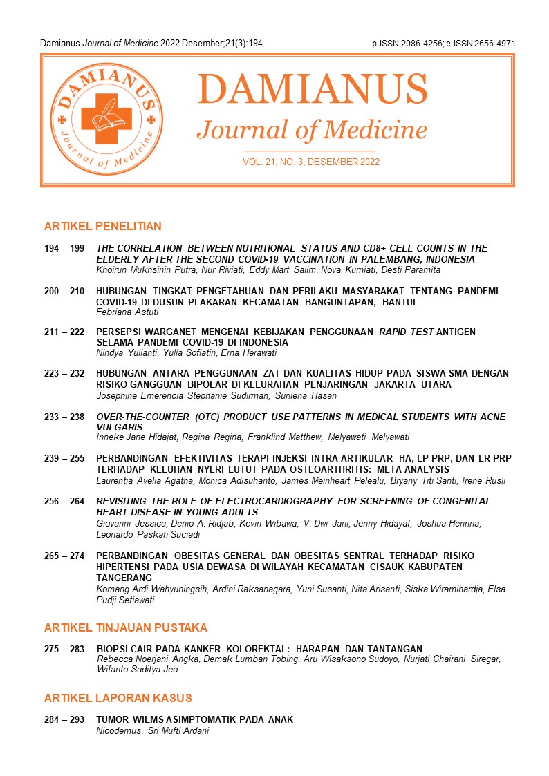
Revisiting the role of electrocardiography for screening of congenital heart disease in young adults
DOI:
https://doi.org/10.25170/djm.v21i3.3558Keywords:
Congenital heart disease, electrocardiography, echocardiography, screeningAbstract
Introduction: Congenital heart disease (CHD) is the most prevalent congenital disability found in newborns. Many cases remain unknown until complications occur, usually in young adults. Transthoracic and Doppler echocardiography are modalities of choice for CHD detection, but these are limited in Indonesia. Electrocardiography (ECG) is a widely available and low-cost test. This study investigated the role of ECG as a screening modality for CHD.
Methods: A cross-sectional study was held at Atma Jaya School of Medicine and Health Science from August to November 2019. Participants were students aged 18 years old or more. Exclusion criteria was previously detected for CHD. Data were collected through history taking, anthropometrics, blood pressure measurement, and 12 leads ECG. ECG results were interpreted independently by two cardiologists to minimize observer bias.
Results: A total of 193 students, 78 male and 115 female, participated. The mean age was 19.22±1.85 years. ECG abnormalities were discovered in 57 (29.5%) participants:15 with crochetage sign, 13 right axis deviation, three left axis deviation, nine right ventricle hypertrophy, and nine left ventricle hypertrophy, six left bundle branch block, and two defective T wave. Further evaluation was done with echocardiography in 20 participants, which resulted in one participant having mitral valve prolapsed (MVP).
Conclusion: ECG could detect the characteristic patterns suggestive of CHD, but ECG alone is insufficient to confirm cardiac structural abnormalities.
Downloads
References
2. Knowles RL, Ridout D, Crowe S, Bull C, Wray J, Tregay J, et al. Ethnic and socioeconomic variation in incidence of congenital heart defects. Archives of Disease in Childhood. 2017 Jun 1;102(6):496–502.
3. Data and Statistics on Congenital Heart Defects | CDC [Internet]. [cited 2021 May 14]. Available from: https://www.cdc.gov/ncbddd/heartdefects/data.html
4. Ismail MT, Hidayati F, Krisdinarti L, Noormanto N, Nugroho S, Wahab AS. Epidemiological Profile of Congenital Heart Disease in a National Referral Hospital. ACI (Acta Cardiologia Indonesiana) [Internet]. 2017 Jan 9 [cited 2021 May 14];1(2). Available from: https://jurnal.ugm.ac.id/jaci/article/view/17811
5. Liberman RF, Getz KD, Lin AE, Higgins CA, Sekhavat S, Markenson GR, et al. Delayed Diagnosis of Critical Congenital Heart Defects: Trends and Associated Factors. Pediatrics. 2014 Aug 1;134(2):e373–81.
6. Benjamin EJ, Muntner P, Alonso A, Bittencourt MS, Callaway CW, Carson AP, et al. Heart Disease and Stroke Statistics-2019 Update: A Report From the American Heart Association. Circulation. 2019 05;139(10):e56–528.
7. Burchill LJ, Huang J, Tretter JT, Khan AM, Crean AM, Veldtman GR, et al. Noninvasive Imaging in Adult Congenital Heart Disease. Circulation Research. 2017 Mar 17;120(6):995–1014.
8. A Case Report of Coarctation of Aorta Presenting as ST Elevation in Anterior Chest Leads of ECG with Severe Left Sided Neck Pain. :4.
9. Rawala MS, Rizvi S. A Rare Case of Adult Congenital Heart Disease: Single Ventricular Chamber with Anomalous Right Coronary Artery. Journal of the American College of Cardiology. 2019 Mar 12;73(9 Supplement 1):2758.
10. Brink a. J., Neill Catherine A. The Electrocardiogram in Congenital Heart Disease. Circulation. 1955 Oct 1;12(4):604–11.
11. Webb Gary, Gatzoulis Michael A. Atrial Septal Defects in the Adult. Circulation. 2006 Oct 10;114(15):1645–53.
12. Ellis JH, Moodie DS, Sterba R, Gill CC. Ventricular septal defect in the adult: Natural and unnatural history. Am Heart J. 1987 Jul;114(1):115–20.
13. Neumayer U. Small ventricular septal defects in adults. Eur Heart J. 1998 Oct;19(10):1573–82.
14. Spicer DE, Hsu HH, Co-Vu J, Anderson RH, Fricker FJ. Ventricular septal defect. Orphanet J Rare Dis. 2014 Dec 29;9(1).
15. Deri A, English K. Educational Series In Congenital Heart Disease: Echocardiographic assessment of left to right shunts: atrial septal defect, ventricular septal defect, atrioventricular septal defect, patent arterial duct. Echo Res Pract. 2018 Mar;5(1):R1–16.
16. Bayar N, Arslan Ş, Köklü E, Cagirci G, Cay S, Erkal Z, et al. The Importance of Electrocardiographic Findings in the Diagnosis of Atrial Septal Defect. Kardiol Pol. 2015 May 20;73(5):331–6.
17. Sabzi F, Faraji R. Adult patent Ductus Arteriosus complicated by endocarditis and hemolytic anemia. Colomb Médica. 2015;46:4.
18. Schneider DJ, Moore JW. Patent Ductus Arteriosus. Circulation. 2006 Oct 24;114(17):1873–82.
19. Sheikh N, Adhikary DK. Coarctation of Aorta. 1. 2014 Jul 6;13(1):56–9.
20. Torok RD. Coarctation of the aorta: Management from infancy to adulthood. World Journal of Cardiology. 2015;7(11):765.
21. Carabello BA, Paulus WJ. Aortic stenosis. The Lancet. 2009 Mar 14;373(9667):956–66.
22. Campbell M, Kauntze R. CONGENITAL AORTIC VALVULAR STENOSIS. Br Heart J. 1953 Apr;15(2):179–94.
23. Heller J, Hagège AA, Besse B, Desnos M, Marie F-N, Guerot C. “Crochetage” (Notch) on R wave in inferior limb leads: A new independent electrocardiographic sign of atrial septal defect. J Am Coll Cardiol. 1996 Mar 15;27(4):877–82.
24. Shen L, Liu J, Li J-K, Xu M, Yuan L, Zhang G-Q, et al. The Significance of Crochetage on the R wave of an Electrocardiogram for the Early Diagnosis of Pediatric Secundum Atrial Septal Defect. Pediatr Cardiol. 2018 Jun;39(5):1031–5.
Downloads
Published
Issue
Section
License
Copyright (c) 2022 Damianus Journal of Medicine

This work is licensed under a Creative Commons Attribution-ShareAlike 4.0 International License.


